Follow Us
Newsletter Sign Up
Copyright © Sarvodaya Hospital 2026. All Rights Reserved.
Disclaimer: The information available on the Sarvodaya Healthcare website, including but not limited to Centres of Excellence, Departments, Treatments, Health Information, Blogs, FAQs, Videos, Graphics, Illustrations, Rare Cases, and other related content, is provided solely for general informational and educational purposes.
The information published on this website is not intended to be a substitute for professional medical advice, consultation, diagnosis, treatment, or prescription. It should not be relied upon for diagnosing or treating any medical condition or for making decisions regarding your health or medical care.
Every individual’s medical condition is unique. We strongly recommend consulting a qualified healthcare professional for appropriate medical assessment, diagnosis, treatment, and advice based on your specific health needs and circumstances.

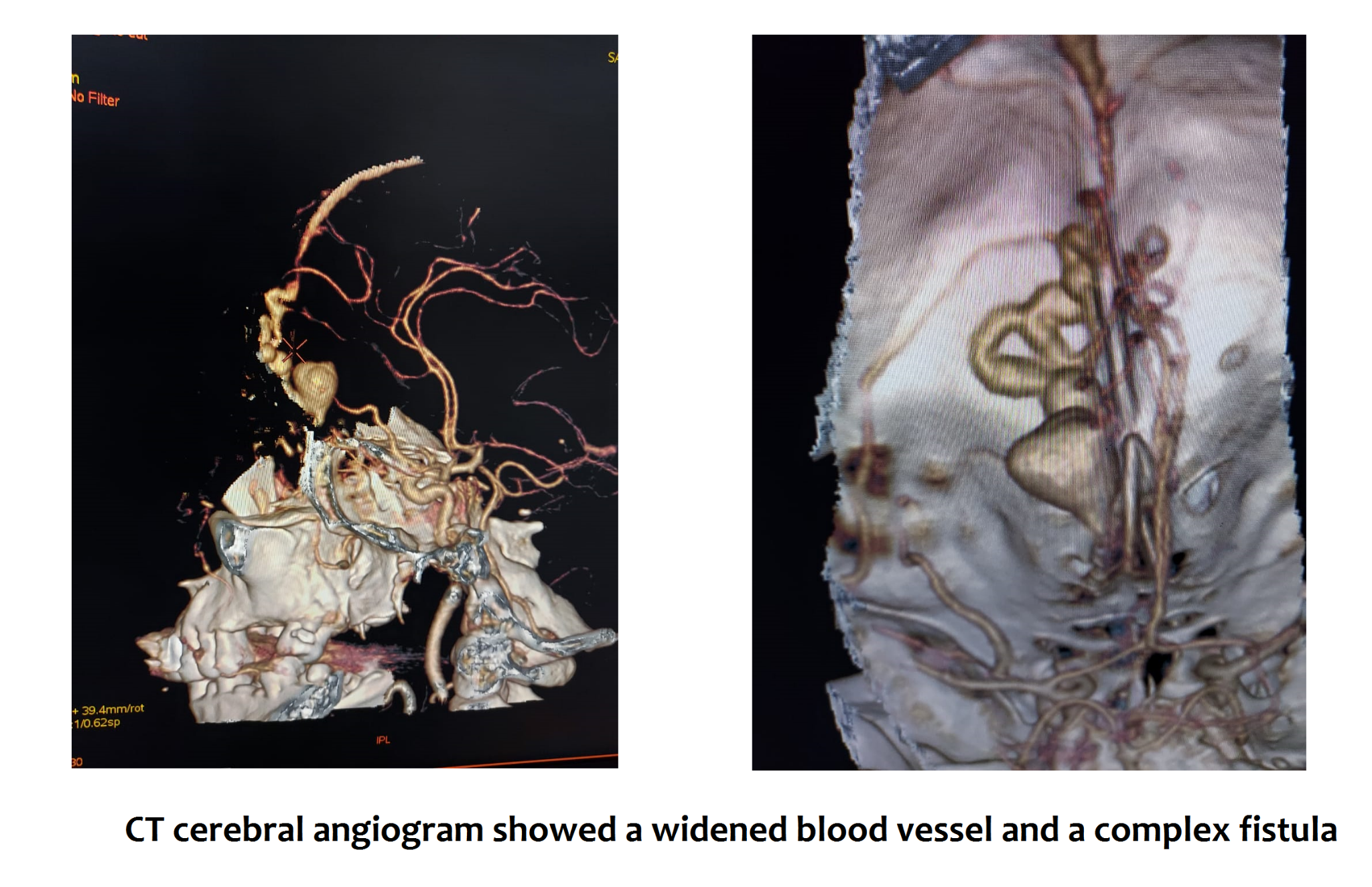
56-year-old male, came with a severe case of Dural Arteriovenous Fistula (an abnormal coiled bundle of arteries and veins) in the brain and a huge Intracranial Haemorrhage (ruptured veins and arteries inside the brain) with a lot of blood leakage. The patient was successfully treated with precise surgical clipping and excision of the Arteriovenous Fistula by our expert neurosurgery team.
“Mr. Suresh Kumar, who had a history of slurry speech, was brought to our emergency department with complaints of headache and multiple episodes of vomiting. On conducting initial investigations via CT scan of the brain, it was found that he had a left frontal Intracerebral Brain Haemorrhage (ruptured veins inside the brain) which further initiated blood leakage to other areas in the brain. A CT cerebral angiogram showed a widened blood vessel and a complex fistula (abnormal coiling of veins and arteries) arising from the left frontal area of the brain. In such critical cases, every second counts, as the blood is constantly leaking to other parts of the brain, thus, without any delay we planned for surgical clipping of ruptured sections and excision of fistula”, said Dr. Kamal Verma, Director, Neurosurgery.
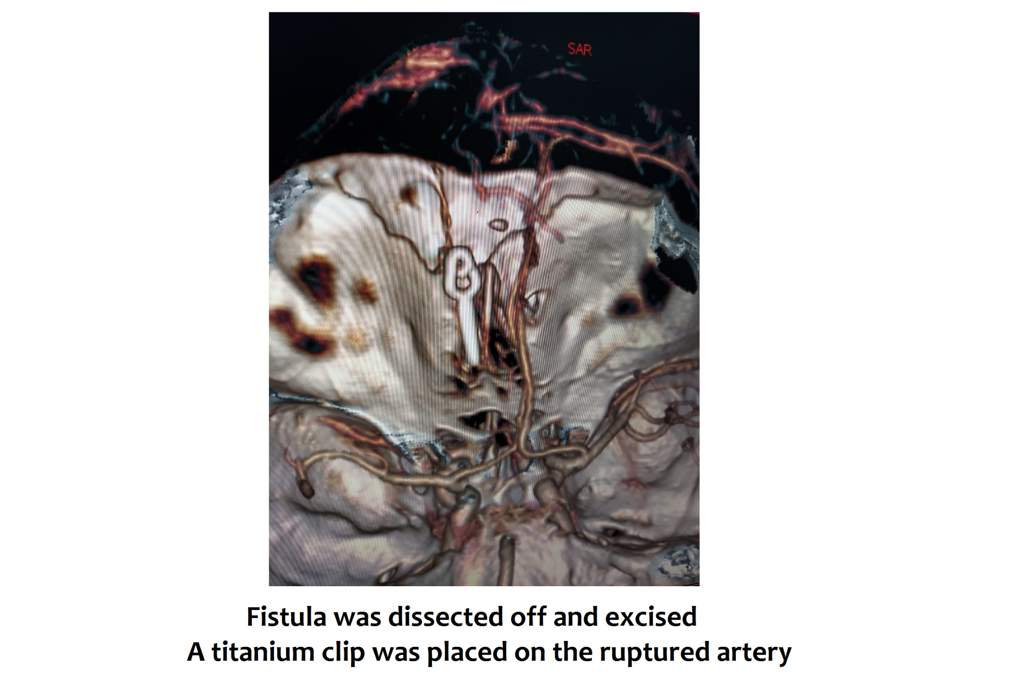
“Considering the critical location of the fistula (near the optic nerve of the eye), we planned for surgery wherein a complex anterior skull base or transnasal approach was taken. In this, we had to surgically open the skull, lift the brain and clip the ruptured blood vessels and excise the fistula which was located near the eyes. We had to be careful during surgery in separating the abnormal vessel and fistula from the normal vessels and the normal brain tissues. Any rupture of the vessel during surgery could have been life-threatening for the patient. The fistula was completely dissected off and excised, a titanium clip was placed on the ruptured artery and the bleed was evacuated”, said Dr. Gaurav Kesari, Consultant, Neurosurgery.
The patient made a good recovery and was taken off the ventilator within 2 days after the surgery. He was completely conscious and was discharged soon.